
Christopher Boone is a math wizard. He can solve incredibly difficult math problems in his head. He is the star of the play “The curious incident of the dog in the nighttime,” a wonderful stage adaptation of the bestselling book by Mark Haddon. Following the applause at the end of the performance, Christopher comes on stage and solves an intricate math problem in his head to the amazement of the audience.

Christopher is autistic, or at least he has a condition that is somewhere on the autism spectrum: a group of neurodevelopmental disorders known as Autism Spectrum Disorders (ASDs). Although he, and certain other people who suffer from these syndromes, can perform incredible feats of memory, mental arithmetic and other cognitive stunts, they also suffer from considerable difficulties when trying to have “normal” social interactions with other people. They are often lonely, obsessive, temperamental and have many other social problems. ASDs are developmental disorders which become apparent during the first few years of life as a child’s behavior becomes increasingly sophisticated. ASDs affect some 60 million people worldwide, and males are much more commonly afflicted than females (about 4 fold). We also know that there are important genetic influences on the development of ASDs, and close relatives of sufferers have an increased risk of developing these disorders. A few individual genes have been demonstrated to have powerful effects on the development of ASDs, but these only account for a few percent of the victims. Clearly, in most instances, there is a complex interaction between environmental and genetic factors that underlies the development of an ASD in a particular patient.
But exactly what is going on in most ASD patients remains a complete mystery. Of course, there are a lots of theories ranging from the completely discredited idea that autism is the result of vaccination, to extremely sophisticated ideas about disruption of the development of the nervous system. A few weeks ago, a new paper appeared in the journal Nature (Reed et al., 2020) supporting the idea that an important factor in the pathogenesis of ASDs was the action of a molecule called interleukin 17a (IL-17a). The study suggested that IL-17a might be helpful in controlling the symptoms exhibited by some ASD patients. By chance, on the very same day, I received an invitation to attend a meeting to discuss the role of IL-17a as a factor promoting diseases like psoriasis. Antibodies such as ixekizumab, which block the effects of IL-17a, seem to be helpful in treating several diseases including psoriasis. Hence, it appears that IL-17a can be helpful or harmful, depending on the circumstances. So, what is going on?
IL-17a is what is known as a proinflammatory cytokine. These molecules are generally small proteins which, as their name implies, are important for regulating inflammatory responses. The cells that produce IL-17a are white blood cells known as T-lymphocytes. A particular subset of these cells, the T-helper 17 (Th17) cells, are an important source of the cytokine. One of the most prominent theories concerning the development of ASDs is that they result from the consequences of the effects of inflammatory cytokines during fetal development when a pregnant mother has an inflammatory reaction due to an infection or some other problem. Because the inflammatory response involves activation of portions of the immune system, this phenomenon is known as Maternal Immune Activation (MIA). Indeed, there is substantial epidemiological evidence suggesting that mothers who have infections or other inflammatory events during pregnancy have offspring with an increased chance of developing ASDs.
Laboratory experiments aimed at determining whether this hypothesis is true have mostly been performed on mice. To model MIA as a risk factor for ASDs, inflammation has been induced in pregnant mice using several approaches. One frequently used protocol is to administer a high single-dose intraperitoneal injection of poly (I:C) to pregnant mice on embryonic day (E) 12.5. This gestational stage resembles the late first trimester in humans, during which viral infections have been correlated with increased incidence of ASDs in the offspring. Poly (I:C) evokes a pro-inflammatory antiviral response in the mother that is similar to the immune response occurring after activation of Toll-like receptor 3 (TLR3) by viral infection. Mimicking MIA during viral infection in this way results in mouse offspring that demonstrate many ASD-like behaviors including defects in social preference, communicative impairments, and repetitive/stereotyped behaviors. Abnormal ultrasonic vocalization observed in these mice is supposed to mirror communication challenges in ASDs, reduced social approach is supposed to mirror impaired social interactions in ASDs, and increased marble-burying behavior is supposed to mirror repetitive behaviors observed in ASDs. MIA also seems to cause changes in the development of the brain and the structure of synapses between individual nerve cells. These may be particularly related to connections between neurons that utilize the inhibitory neurotransmitter GABA. In ASDs, the degree of inhibitory GABA-mediated neurotransmission in the brain may change relative to the degree of excitatory glutamate-mediated neurotransmission, meaning that some brain networks are hyperexcited. In this context, several recent investigations have suggested that imbalances in the GABAergic neurotransmission system may be implicated in the development of ASDs as well as several other related neurodevelopmental disorders, including Fragile X Syndrome and Rett Syndrome.
Of course, whether such mouse behaviors really have anything to do with human autism is a matter for discussion. Mice are not humans. ASDs are human diseases and mice may not be able to produce behaviors that truly mimic the human disease. However, this is the state of the art. A great deal of time, effort and expense has been invested into establishing these mouse models as an experimental system for studying human autism. Unfortunately, modeling human cognitive diseases in mice has not proved to be a very useful approach to finding cures for human diseases—so we will have to wait and see whether studies of autism in mice can buck this trend.

As mentioned above, poly (I:C) activates a receptor on many immune cells called TLR3. One consequence of this activation is that the cells start to make a large number of inflammatory cytokines. Although inflammatory cytokines have important actions in coordinating inflammatory responses, which are important in maintaining tissue homeostasis in the face of injury or infection, we also know that when these molecules are present in abnormally large amounts they can wreak havoc, disrupting the functions of many tissues including the brain. Consider, for example, the cutting-edge form of cancer treatment known as CAR-T cell therapy. This cancer treatment involves using specially engineered T-lymphocytes to target and destroy cancer cells. Killing the cancer targeted by CAR-T cells is mediated through the release of inflammatory cytokines among other mechanisms. In some individuals this happens to excess, producing what is called a “cytokine storm.” The clinical manifestation of this syndrome resembles sepsis, with symptoms such as high fever, fatigue, myalgia, nausea, capillary leakages, tachycardia and other cardiac dysfunction, liver failure, and kidney impairment. Neurological symptoms such as seizures can occur and even death. Clearly, then, a large rapid systemic release of inflammatory cytokines can be extremely toxic.
In the case of a pregnant mouse treated with an injection of poly (I:C), the production of inflammatory cytokines may not be quite so dramatic but it is still sufficient to produce increases in the blood levels of many of these molecules and clear behavioral consequences in offspring. Because many different cytokines are produced following poly (I:C) treatment, it was of interest to determine which ones in particular were responsible for the autism-like behavioral changes observed in offspring. Further experiments demonstrated that interfering with IL-17a and its ability to activate its receptor prevented the behavioral abnormalities and structural brain changes in young mice from MIA mothers. On the other hand, injection of 1L-17a into the brains of young mice produced the same changes as observed in the offspring of MIA mothers. So, it was concluded that 1L-17a signaling was responsible for the autism-like effects of MIA mothers on their offspring.
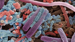
Such results are of interest but turned out to have several surprising consequences. The first of these concerns the question of how MIA normally occurs in the mothers of autistic children. In other words, what normally promotes such events in pregnant mothers? Over the last decade, there has been an increasing realization that animals like humans don’t really exist as single organisms. We actually go through life living a symbiotic existence with trillions of single-cell organisms such as bacteria, archaea, protists, fungi and viruses. These organisms live primarily in our gastrointestinal tract as well as other tissues including the skin, mammary glands, placenta, seminal fluid, uterus, ovarian follicles, lung, saliva, oral mucosa, conjunctiva and biliary tract. From a genetic point of view, this vast repository of microorganisms is known as the microbiome. The microbiome metabolizes many substances contained in food and other sources, and these metabolites play an important role in signaling to tissues throughout the body. Surprisingly, changes in the microbiome can even exert influences on the functioning of the central nervous system. Moreover, the microbiome has many influences on the immune system, particularly in “training” the immune system of infants to develop immunity and in the regulation of the immune response. Effects of the microbiome on the brain may even extend to having profound influences on the progression of brain diseases. Autism is no exception to this. In addition to immune dysfunction, ASDs in humans have also been associated with changes in the population of the gut microbiome (dysbiosis) and gastrointestinal inflammation. The occurrence of these phenomena has led to increasing speculation about a role for the microbiome in ASDs. Here again, animal studies have attempted to provide a mechanistic understanding of how the microbiota could have a role in the generation of ASDs. In these studies, the complement of gut microbes was specifically altered and then the propensity of mice to develop ASD-like symptomology was assessed under different circumstances. For example, germ-free mice exhibited ASD-like deficits in social behavior and increased repetitive behavior, suggesting that appropriate composition of the microbiota is required for normal social development. As would be predicted, such deficits could be remedied by supplying different types of gut microbes to germ-free mice.
Administration of a single bacterial strain, either Bacteroides fragilis or Lactobacillus reuteri, was found to reverse many of the behavioral and gastrointestinal changes reported in both human studies and animal models of ASD. On the other hand, transplantation of gut microbiota from human donors with ASDs into germ-free mice revealed that colonization with microbiota from people with ASDs was sufficient to induce autistic behaviors in recipient mice. Moreover, the presence of some gut microbial communities that are susceptible to vancomycin and that promote a pro-inflammatory status has been associated with ASDs. Finally, administration of probiotics (usually live microbial cultures that impart a health benefit to the host) or prebiotics (non-digestible foodstuffs, including fiber, which convey a beneficial effect for the host or microbiota) has been shown to modulate the social behavior of animals. These studies, suggesting a potential role of diet on the occurrence of ASDs, are very provocative and, if they could be translated to patients with ASD, might be extremely influential from the therapeutic point of view.
Experiments performed on mice again attempted to further define the mechanisms that underlie these effects. Experimenters determined that alterations to the ecology of the gut microbiome altered the ability of MIA treatments to produce Th17 cells and ASD-associated symptoms in experimental animals. Hence, the data strongly support a model in which MIA is associated with autism in offspring, particularly when the production of IL-17a is primed through the composition of the gut microbiota. Women with gut microbial communities that promote excessive Th17 cell differentiation may therefore be more likely to bear children with ASDs in the event of pathological inflammation during pregnancy. The data also suggest that a reduction of IL-17a during pregnancy could reduce the likelihood of the occurrence of autism.
However, things haven’t turned out to be quite so straightforward, and the story has recently been shown to have an interesting twist. A surprising but reproducible observation has been made that, although inflammation in pregnant mothers seems to be a risk factor for the development of ASDs by their offspring, some autistic children actually exhibit temporary but substantial improvements in their behavioral symptoms during episodes of fever, a sign of systemic inflammation. In their recent paper, Reed et al. investigated the mechanism of this effect. In these experiments, the authors used several mouse models of autism including the offspring of MIA mice, as described above, and also genetic mouse models for ASDs, in which mice express mutations in ASD risk genes (including Cntnap2, Fmr1, and Shank3). As with MIA offspring, these mouse mutants exhibit behavioral abnormalities, such as deficits in social interactions. Reed et al. induced fever in these animals by giving them lipopolysaccharide (LPS), which—like poly (I:C)—is an inducer of the inflammatory cytokine response, in this case through the activation of Toll-like receptor 4 (TLR4). The offspring of MIA mice exhibited the well-known lack of sociability. However when assessed 4 hours after an LPS injection, their sociability rating was much improved and they behaved more or less like ordinary mice. Abnormal behavior was apparent again when the mice were tested 72 hours after the LPS injection, indicating the transient nature of the improvement. Moreover, if a fever was induced in the offspring of MIA mice by using a different procedure, no improvement in the behavior of the mice was observed, indicating that it wasn’t the fever response per se that was responsible for the LPS-induced improvement. The effects of LPS were also apparent when the activity of neurons in the brain was examined. It is known that MIA offspring suffer from a disorganization of certain cortical neurons. The activity of neurons in this region of the cortex is increased in MIA offspring and in the mutant mouse lines. If this increased activity was artificially reduced using a technique known as optogenetics, then sociability in the MIA offspring and some of the mutant mice was increased. These results suggest that LPS-induced reduction of the activity of a specific population of cortical neurons is responsible for the improvement of sociability behavior. This of course raises the question as to exactly how the effects of LPS on these neurons are produced? A search for the signaling molecule that was responsible for these effects of LPS produced a surprising result. The intermediary molecule responsible for the observed effects was once again IL-17a! The authors of the paper observed that receptors for IL-17a were expressed in the appropriate cortical neurons, that direct injection of IL-17a into the cortex reduced the activity of these neurons and improved sociability. Furthermore, they demonstrated that an injection of an antibody against IL-17a into mice blocked the ability of LPS to improve sociability . All of these studies point to the conclusion that IL-17a is an important regulator of the appearance of ASDs, at least in some people. IL-17a is a key intermediate in the induction of mouse ASD symptoms in the offspring of MIA mice and, in contrast to this, it actually helps to suppress these same symptoms in adult ASD mice born to MIA mothers.
These studies highlight the intricate actions of inflammatory cytokines in the pathogenesis of ASDs and suggest that manipulating IL-17a in different ways at different stages of the disease might be beneficial in the treatment of ASDs. The experiments described above have all concentrated on measuring specific ASD-related effects in mice. But, of course, the role of inflammatory cytokines is not just to control the symptoms of ASDs—and IL-17a is a good example of this. As it turns out, IL-17a is already a therapeutic target for other disease indications. For example, consider the inflammatory skin disease known as psoriasis which is often associated with a type of inflammatory arthritis. It turns out that the inflammatory events that drive these syndromes are also critically dependent on IL-17a. A great deal of research has demonstrated the presence of IL-17a and its receptor in skin lesions from psoriasis patients. What is more, interference with IL-17a by neutralizing its action is a very helpful way of treating psoriasis or associated arthritis. There are several monoclonal neutralizing antibodies against IL-17a, such as ixekizumab, that are effective in patients with the disease. Moreover, IL-17a has been indicated as playing a role in a large number of other diseases which are thought to have an inflammatory component in their progression, even including things such as Alzheimer’s disease. Hence, even though the effects of IL-17a in ASDs might be beneficial or detrimental, just giving patients IL-17a or an antibody against it is unlikely to work as a therapy for the disease, given all of the other effects of this cytokine. Nevertheless, that does not rule out the possibility that the IL-17a signaling pathway could be targeted in some way to therapeutic advantage. A clear understanding of the molecular signaling pathways involved in expression of a disease is certainly an important start in understanding what kind of therapeutic agents may be of use.
Studies that have revealed the potential effects of IL-17a in the pathogenesis of ASDs and the role of the microbiome in this regard are a great illustration of how different biological mediators can play varying roles under a variety of different circumstances, and why targeting these things therapeutically is always likely to be a challenging but interesting opportunity. These studies have been very illuminating. It will be of great interest to see if they can be translated to human patients in the not-too-distant future.
IL-17a promotes sociability in mouse models of neurodevelopmental disorders.
Reed MD, Yim YS, Wimmer RD, Kim H, Ryu C, Welch GM, Andina M, King HO, Waisman A, Halassa MM, Huh JR, Choi GB.
Nature. 2020 Jan;577(7789):249-253. doi: 10.1038/s41586-019-1843-6.